Calcaneal Slide Osteotomy Technique
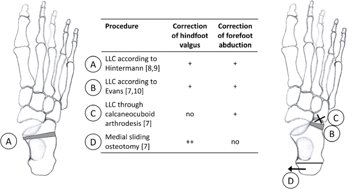
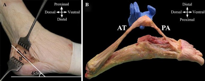
The medial slide calcaneal osteotomy alters the pull of the gastrocnemius soleus muscle group slightly medial to the axis of the subtalar joint.
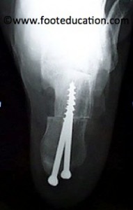
Calcaneal slide osteotomy technique. Obtains focused history and physical. Surgeons can then achieve fixation using the 7 0 mm compression ft screws or the 6 5 compression pt screws. Specific techniques in a calcaneal osteotomy an incision is made on the outer or lateral side of the foot. The literature shows a high rate of hardware prominence with screws leading to subsequent removal of hardware.
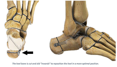
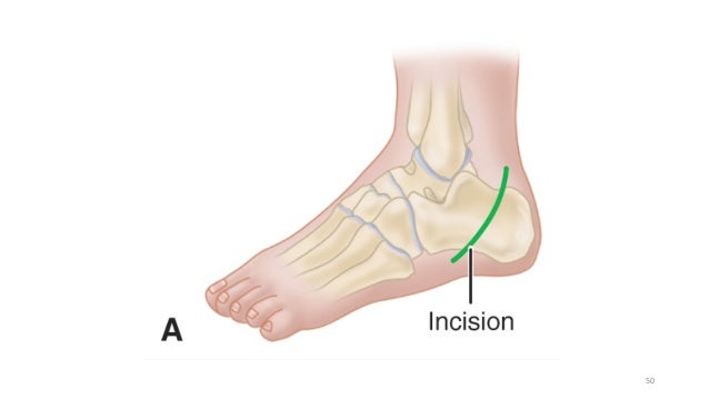
The lateral calcaneus cortex is exposed using a lateral incision. The posterior osteotomy fragment is manually mobilized and shifted laterally. Plantar fascia release and calcaneal slide osteotomy are often components of the surgical management for cavovarus deformities of the foot. The purpose of a calcaneal osteotomy is to shift the heel bone towards the inside medial or outside lateral.
After the bone is cut it is moved to the desired location and fixed in place. The medializing calcaneal osteotomy is a frequently performed procedure usually done in conjunction with a flexor tendon transfer as part of a flatfoot correction surgery. Most often surgical implants such as screws hold the bones together and support healing. The calcaneal slide osteotomy is a common procedure used for the surgical correction of heel varus and valgus deformities.
A calcaneal osteotomy is a common technique used to treat stage ii flatfoot deformity. In this setting plantar fascia release has traditionally been performed through an incision over the medial calcaneal tuberosity and the calcaneal osteotomy through a lateral incision. By sliding the posterior tuber of the calcaneus 1 cm medially the mechanical axis of the limb is medialized thereby decreasing the stress on the posterior tibial tendon s mechanics to invert the hindfoot. A calcaneal osteotomy is a bone cut osteotomy that a surgeon makes across the heel bone calcaneus.
Intermediate evaluation and management. Arthrex s recently launched minimally invasive surgery platform allows surgeons to perform this osteotomy through a tiny incision. The osteotomy is performed through an oscillating saw. This effectively places the achilles tendon medially and increases the varus pull on the hindfoot while correcting the rearfoot valgus alignment.
Some patients have a heel bone that is shifted or tilted too far to the inside or outside of their shin bone. Deirdre ryan los angeles robert kay los angeles children s hospital los angeles approved technique video 0 technique steps 0.


